단백질 인산화효소는 세포주기 조절과 분화를 포함해 다양한 세포 기능에 관여합니다. Bcr/Abl이나 K-Ras와 같은 발암성 인산화효소에 대한 연구는 종양학을 발전시키고 항암치료 전략으로서 표적화 약물과 맞춤의학의 등장을 견인했습니다.
RTKs/Growth Factor Receptors• EGFR, mutant EGFR
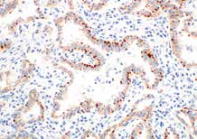
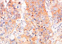
ROS1 (D4D6®) Rabbit mAb #3287: IHC analysis of paraffin-embedded human lung carcinoma using #3287. |
Intracellular Kinases• B-Raf
Ras (E8N8L) XP® Rabbit mAb #67648: IHC analysis of paraffin-embedded human non-small cell lung carcinoma using #67648. |
Kinase Inhibition Readouts• Phospho-Akt
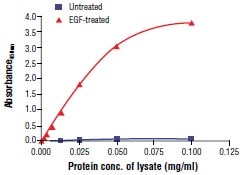
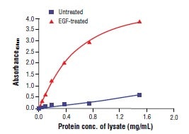
FastScan™ Phospho-p44/42 MAPK (Erk1/2) MAPK (Thr202/Tyr204) ELISA Kit #42173 |
세포주기 관문(checkpoint)과 DDR 경로는 협력하여 유전체(genome)를 보전합니다. 둘 중 하나라도 불안정하면 통제되지 않는 세포 증식과 돌연변이 축적으로 인해 종양 발생으로 이어질 수 있습니다. 이들 경로에서 결손을 표적화 함으로써 여러 암 유형에 대한 차세대 치료제를 개발할 수 있습니다.
Cell Cycle Regulation• Phospho-Rb EGFR
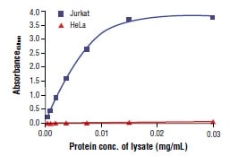
FastScan™ Phospho-Rb (Ser807/811) ELISA Kit #10754 |
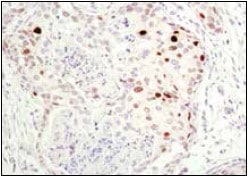
DNA Damage Response (DDR)• Phospho-p53/p53, MDM2
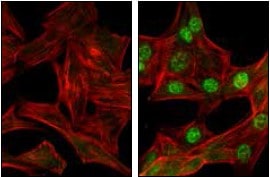
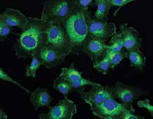
Phospho-Chk1 (Ser345) (133D3) Rabbit mAb #2348: Confocal IF analysis of C2C12 cells, untreated (left) or UV-treated (right), using #2348 (green). Actin filaments have been labeled with |
Senescence• p16 INK4A
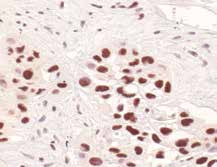
p21 Waf1/Cip1 (12D1) Rabbit mAb #2947: IHC analysis of paraffin-embedded human breast carcinoma using #2947 in the presence of control peptide. |
암세포 대사의 특징을 기술하는 것으로 시작된 암 대사 연구는 암세포가 대사학적으로 어떻게 재프로그래밍되어 이동, 침범, 전이에 영향을 미치는지를 이해하는 수준까지 발전했습니다. 대사경로를 표적화 하는 항암치료제는 암 성장을 억제하고 여러 가지 병용요법을 선택할 수 있게 해줍니다.
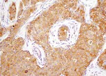
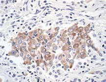
Glucose Metabolism• Phospho-PKM2/PKM2
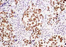
PKM2 (D78A4) XP® Rabbit mAb #4053: IHC analysis of paraffin-embedded human lung carcinoma using #4053. |
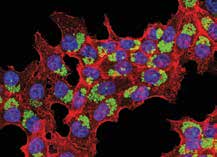
Autophagy• LC3B
LC3B (E5Q2K) Mouse mAb #83506: Confocal IF analysis of HCT 116 cells treated with Chloroquine #14774 (50 μM, overnight) using #83506 (green). Actin filaments were labeled with β-Actin (13E5) Rabbit mAb #4970 (red) and nuclei were labeled with DAPI #4083 (blue). |
Fatty Acid and Amino Acid Metabolism• Phospho-Acetyl-CoA Carboxylase/
Phospho-Acetyl-CoA Carboxylase (Ser79) (D7D11) Rabbit mAb #11818: IHC analysis of paraffin-embedded human breast carcinoma using #11818. |
상피-중간엽 전이(EMT)는 암이 악성으로 진행되는 데에 핵심적 역할을 해서 전이와 치료 내성 발생을 촉진한다고 알려진 세포 내 프로그램입니다. EMT 활성화를 표적으로 한 새로운 항암제는 암이 전신으로 전이되는 것을 억제하고 치료 경과를 개선할 수 있습니다.
Transcription Factors/Co-activators• Phospho-STAT3 ELISA
PathScan® Phospho-Stat3 (Tyr705) Sandwich ELISA Kit #7300 |
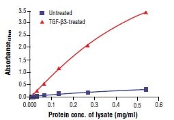
Signaling Pathways• Phospho-SMAD2/3 ELISA, TGF-β
FastScan™ Phospho-Smad2 (Ser465/467) ELISA |
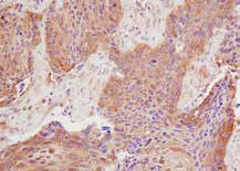
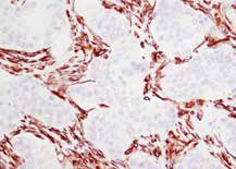
Key Biomarkers• Vimentin
Vimentin (D21H3) XP® Rabbit mAb #5741: IHC analysis of paraffin-embedded human breast carcinoma using #5741. |
세포사멸 회피는 암의 대표적 성질입니다. 암세포의 사멸을 촉발시키는 세포사멸 경로를 표적화 하는 것은 한 가지 암 유형에 국한하지 않고 널리 활용가능한 항암제의 개발을 기대할 만한 유망한 전략입니다.
Intrinsic Pathway• Bcl-2, Bcl-xL, Mcl-1
Bcl-xL (54H6) Rabbit mAb #2764: IHC analysis of paraffin-embedded human lung carcinoma, using #2764. |
Extrinsic Pathway• TRAIL, DR4, DR5
DR5 (D4E9) XP® Rabbit mAb #8074: Confocal IF analysis of HT-1080 cells using #8074 (green). Blue pseudocolor = DRAQ5® #4084 (fluorescent DNA dye). |
Caspase Activators• Smac/Diablo
Survivin (71G4B7) Rabbit mAb #2808: IHC analysis of paraffin-embedded human transitional epithelial carcinoma of the bladder using #2808. |
비정상 후성유전학적 반응과 유전자 돌연변이 모두 종양생성에 기여하지만, 현재 대부분의 항암제는 유전자 이상에 표적화 하는 것에 집중하고 있습니다. 신생 세포를 정상세포와 비슷하게 행동하도록 재프로그래밍하는 후성유전학적 약물은 기존 치료 전략의 대안이 될 수 있으며 다른 항암제와의 복합요법으로서 가장 큰 효과를 발휘할 것으로 기대됩니다.
Methylation and Demethylation Inhibitors• Histones Regulators: Ezh2, LSD1, H3K27me3
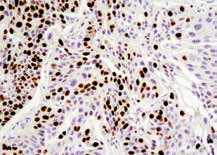
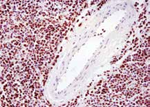
Ezh2 (D2C9) XP® Rabbit mAb #5246: IHC analysis of paraffin-embedded human lymphoma using #5246. |
Acetylation and Deacetylation Inhibitor
• Histones Regulators: CBP, p300, H3K27ac, HDACs
p300 (D8Z4E) Rabbit mAb #86377: IHC analysis of paraffin-embedded human squamous cell lung carcinoma using #86377. |
Histone Mutations
• Histones: H3K27M, H3K36M, H3K9M,
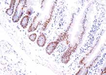
Histone H3 (K9M Mutant Specific) (E4N7V) Rabbit mAb #54905: IHC analysis of paraffin-embedded histone H3 K9M mutant mouse small intestine using #54905. (Tissue courtesy of Dr. Aaron Huebner, Hochedlinger Lab at Massachusetts General Hospital, Boston, MA.) |
종양의 성장과 생존은 악성세포 뿐만 아니라 종양미세환경(TME)을 구성하는 내피세포, 기질 섬유아세포, 면역세포로부터의 신호에 의해 좌우됩니다. 내피세포 및 섬유아세포와의 복잡한 상호작용 뿐만 아니라 세포독성 T 세포에 의한 종양 침투 정도를 바탕으로 임상적 경과를 예측하고 조정할 수 있습니다.
Immune Cell Infiltration• CD3
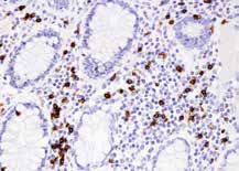
CD8α (D8A8Y) Rabbit mAb #85336: IHC analysis of paraffin-embedded human Crohn’s diseased colon using #85336. |
Immune Cell Function• FoxP3
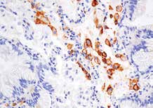
CD86 (E2G8P) Rabbit mAb #91882: IHC analysis of paraffin-embedded human lung denocarcinoma using #91882 performed on the Leica BOND Rx. |
Therapeutic Targets• CD47
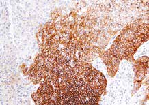
CD47 (D3O7P) Rabbit mAb #63000: IHC analysis of paraffin-embedded human urothelial carcinoma using #63000 performed on the Leica BOND Rx. |
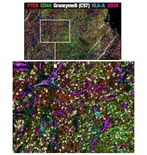
Multiplex IHC CompatibilityGold-Standard IHC Validation Enables Multiplex Discovery
Multiplex IHC analysis of paraffin-embedded malignant melanoma on the CODEX platform using Granzyme B (D6E9W) Rabbit mAb #46890, shows the widespread labeling of infiltrating immune cells in this sample, along with other markers. |
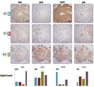
Proteomic and Genomic Correlation using Nanostring TechnologyReliably Correlate Protein Expression with Genomic Data and IHC
Serial sections of each NSCLC specimen were stained with antibodies for each of the noted proteins. Tumor Ras G12C genotype of each sample (A–C) obtained from SNV analysis is indicated (+/-) on the left. An additional single serial section was simultaneously stained with DNA-barcoded antibodies. Normalized digital counts from |
PD-1, PD-L1, CTLA-4와 같은 면역관문을 차단하는 T 세포-표적성 면역조절제는 항암치료의 접근 방식에 혁신을 초래했습니다. 이 치료법은 후기 병기의 일부 환자에서 유의미하게 경과를 개선하고 있습니다.
Checkpoint Therapy Targets• PD-1
TIGIT (E5Y1W) XP® Rabbit mAb #99567: IHC analysis of paraffin-embedded human non-small cell lung carcinoma using #99567. |
T Cell Exhaustion• PD-1
Tox/Tox2 (E6I3Q) Rabbit mAb #73758: IHC analysis of paraffin-embedded human colon carcinoma using #73758 performed on the Leica® BOND™ Rx. |
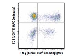
T Cell Activity• IFN-γ
IFN-γ (D3H2) XP® Rabbit mAb (Alexa Fluor® 488 Conjugate) #12942: Flow cytometric analysis of human peripheral blood mononuclear cells treated with TPA, Ionomycin and Brefeldin |
선천적 면역계를 활성화하면 암세포가 촉발하는 면역 회피 경로를 막을 수 있습니다. 이 기전을 심층 이해한다면 다른 치료 전략과 병용하여 장기적으로 임상적 혜택을 유도할 새로운 면역항암제를 개발할 수 있습니다.
Inflammasome• NLRP3
IL-1β (3A6) Mouse mAb #12242: IHC analysis of paraffin-embedded human large intestine (ulcerative chronic colitis of the rectum) using #12242. |
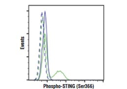
STING• cGAS
Phospho-STING (Ser366) (E9A9K) Rabbit mAb #50907: Flow cytometric analysis of TPA differentiated THP-1 cells that have been activated to phosphorylate STING (green line) vs unactivated (blue). Isotype controls are shown by dashed lines. |
TLR• Phospho-IκBα/IκBα
NF-κB p65 (D14E12) XP® Rabbit mAb #8242: IHC analysis of paraffin-embedded human chronic cholecystitis using #8242. |
면역세포 활성화 측정을 통해 새로운 면역항암제의 유효성 결정을 위한 선천적 면역계와 후천성 면역계의 활성화 정도를 평가할 수 있습니다.
T Cell Signaling• Phospho-SLP-76 (Ser376)
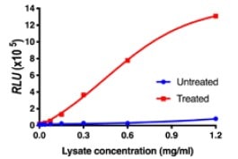
PathScan® Phospho-SLP-76 (Ser376) Sandwich |
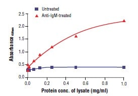
B Cell Signaling• Phospho-Syk (Tyr525/526)
PathScan® Phospho-Btk (Tyr223) Sandwich ELISA Kit #23843 |
Cytokine Response• Phospho-STAT3 (Tyr705)
FastScan™ Phospho-Stat1 (Tyr701) ELISA Kit #40716 |
적응세포치료법은 종양침투성 림프구를 활용하거나 새로운 T 세포 수용체(TCR)나 키메라 항원 수용체(CAR)를 발현하도록 유전자 조작된 T 세포를 활용해 종양세포를 박멸하는 면역치료 전략입니다. 이 치료전략은 항-종양 효과 개선, 독성 감소, 동종세포(allogenic) 이용가능성 파악을 목표로 하여 개발되고 있습니다.
Monitoring CAR-T Target Expression• TNFRSF17/BCMA
TNFRSF17/BCMA (E6D7B) Rabbit mAb #88183: IHC analysis of paraffin-embedded human normal colon using #88183. |
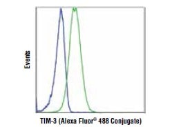
T Cell Phenotyping• CD4
TIM-3 (D5D5R™) XP® Rabbit mAb (Alexa Fluor® 488 Conjugate) #54669: Flow cytometric analysis of primary CD4+ T cells (green, positive) and Jurkat cells (blue, negative). |
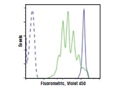
T Cell Functional Analysis• Cytotoxicity assay
Cell Proliferation Tracer Kit (Fluorometric, Violet 450) #48444: Live human peripheral blood mononuclear cells were labeled with the kit comparing treatment to induce cellular division (green) vs. untreated (solid blue line). Unstained cells were used as a control (dashed line). Multiple peaks equate to multiple rounds |
세포 생존력이란 검체 중 건강한 세포의 백분율을 의미합니다. 세포 생존력의 생물학적 측정치를 이용하여 암세포의 증식을 억제하거나 세포사멸을 촉진하도록 개발된 치료제의 안전성과 유효성을 평가합니다.
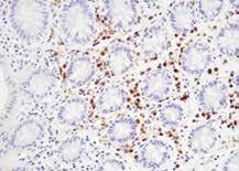
Proliferation• Ki-67
Ki-67 (8D5) Mouse mAb #9449: IHC analysis of paraffin-embedded human breast carcinoma using #9449. |
Cell Death• Apoptosis: Cleaved Caspase-3 ELISA, Cleaved
PathScan® Phospho-RIP (Ser166) Chemiluminescent Sandwich ELISA Kit #88918 |
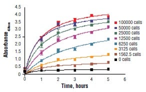
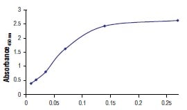
Viability• XTT Cell Viability Assay
XTT Cell Viability Kit #9095: C2C12 cells were seeded at varying density in a 96-well plate and incubated overnight. The XTT assay solution was added to the plate and cells were incubated. The absorbance at 450 nm was measured at 1.0, 2.0, 3.0, 4.0, and 5.0 hours. |