Home » 제품안내 » 이달의 추천제품 » [Cayman] Cell Death Mechanisms & Detection Tools – Apoptosis
[Cayman] Cell Death Mechanisms & Detection Tools – Apoptosis
APOPTOSIS
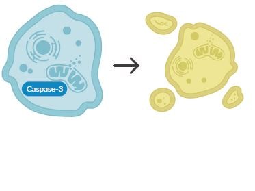
Apoptosis는 다세포 생물체에서 볼 수 있는 Programmed cell death의 일종으로 ATP-dependent하며 치밀한 유전적 프로그램을 따르는 자발적 세포제거 과정입니다. 이 과정은 세포질 수축, DNA fragmentation, Nuclear condensation, 세포막의 변화, 세포사멸체 형성, 그리고 최종적으로 Macrophages phagocytose가 일어나면서 종료됩니다.
Activation of apoptosis signaling cascades
Pannexin channel “find me” signal release
Phosphatidylserine (PS) exposure
Mitochondria outer membrane permeability
Release of cytochrome c from mitochondria
Activation of initiator and effector caspases
Cleavage of specific caspase substrates
Chromatin condensation and DNA fragmentation
Membrane blebbing
Formation of apoptotic bodies
Simultaneous Detection of Multiple Changes in Apoptosis Pathways
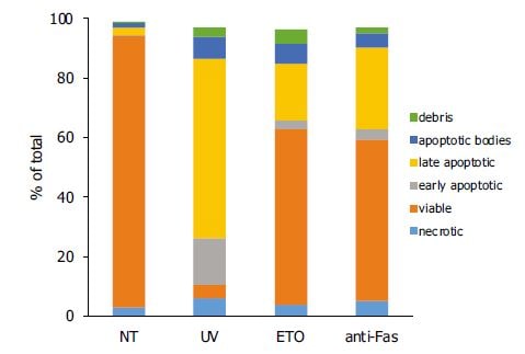
세포 사멸은 다양한 경로로 발생할 수 있으므로 in vitro에서 multiple apoptosis marker를 test하여 apoptosis mechanism을 확인해야 합니다. Cayman에서는 이와 관련된 제품들을 제공하고 있습니다.
Cell populations induced to undergo apoptosis by various stimuli: Untreated (NT); Ultraviolet light (UV); Etoposide (ETO); Anti-Fas CH-11
Contains DAPI – DNA stain measures membrane permeability Annexin V – PS exposure and loss of membrane asymmetry TMRE – Mitochondrial membrane potential TO-PRO®-3 – Identifies active pannexin channels
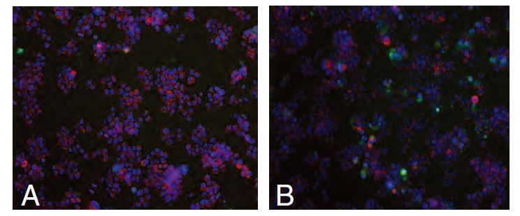
A) Untreated. B) RAW 264.7 cells stimulated to undergo apoptosis by UV concurrently decrease TMRE staining and increase Annexin V FITC staining.
Contains Hoechst 33342 – Demonstrates nuclear morphology Annexin V – PS exposure and loss of membrane asymmetry TMRE – Mitochondrial membrane potential RedDot™2 – DNA stain measures membrane permeability
Pro- and Anti-Apoptotic Signaling Pathways
The pro- and anti-apoptotic Bcl-2 family of proteins regulates commitment to cell death through controlling mitochondrial integrity. Cleavage or oligomerization as well as the abundance of these proteins can mark apoptosis.
PS migrates to the outer plasma membrane in apoptosis, causing a loss of membrane asymmetry. Annexin V binds to exposed PS and can be paired with a membrane-impermeable dye to distinguish between intact, apoptotic, and necrotic cells.
Mitochondrial outer membrane permeabilization causes a decrease in transmembrane potential (Δψm) and the release of cytochrome c. Δψm is assessed using positively charged dyes, such as JC-1 and TMRE, that accumulate inside active mitochondria.
Release of cytochrome c from the mitochondria promotes caspase-9 activation via Apaf-1, which then activates caspases-3 and -7. Caspase activation can be confirmed by detecting cleaved PARP1 or fluorogenic substrates.
The condensation of chromatin is accompanied by the hydrolysis of nuclear DNA into a ladder of fragments that can be detected by gel electrophoresis. The 3’ ends of DNA fragments can also be labeled with deoxyuridine. Cell-permeable Hoechst 33342 is often used to identify nuclear condensation by microscopic analysis.