Quality & Reproducibility
 |
Reproducibility in your experiments is not a matter of chance. It is a matter of science. CST products are developed, tested, and rigorously validated across multiple applications by tenured CST scientists who understand the underlying biology. Over 99.5% of CST recombinant monoclonal antibodies are manufactured in-house, providing complete control over our supply chain and providing lot-to-lot consistency for the lifetime of your project. |
Assay Support & Flexibility
 |
Your assay is only as good as your antibody is specific. CST antibodies and ready-to-use ELISA and cellular assay kits are developed with this in mind. They are designed to seamlessly fit into your assay workflow and instantly answer key questions. CST subject matter experts are available to help identify the best readout and clone to effectively assess your therapeutic efficacy and safety. |
Companion Reagents
 |
CST offers a wide selection of epitope-tagged and control antibodies, secondary antibodies, detection reagents, and experimental controls, as well as standard buffers and other reagents required to complete your experimental workflow. |
Services & Customization
 |
Sometimes the fastest way to move your discovery forward is to have someone else prepare the tools you need. CST provides custom solutions that meet your specific research challenges, freeing up your time to focus on the science. |
| 서비스와 고객 맞춤 제안 | 개요 |
| 캐리어가 필요 없는 맞춤형 제형 (Carrier-free and Customized Formulations) | 대량처리(High Throughput) ELISA 분석, 멀티플렉스 IHC, 유세포분석법 (Flow Cytometry) IHC 자동염색기기(Autostainers), 고성능 대량 스크리닝 및 분석(High Content Screening and Analysis) 등 모든 분석법과 플랫폼에 이상적입니다. |
| 컨주게이션 항체 맞춤 제작 (Custom Antibody Conjugation) | CST의 내부 전문가가 형광단(fluorophore), 비오틴, 효소 등 의뢰된 분석에 필요한 컨주게이션 항체를 직접 제작합니다. |
| 프로테오믹스 분석 서비스 (Proteomics Analytical Services) | CST 전문가 팀이 의뢰된 검체의 정량적 및 정성적 단백 프로파일 분석을 실시합니다. 프로테옴 (Proteome)을 규명하고 처치 전과 후의 번역 후 변형(Posttranslational Modifications)을 확인합니다. |
| 벌크 배송과 로트 예약 (Bulk Quantities and Lot Reservation) | CST가 파트너로 함께 하면 단일 로트 예약이나 대용량 주문이 가능하므로 잠재적 공급 문제가 없어집니다. |
| 펩타이드 맞춤 제작과 관리 (Custom Peptides and Controls) | CST는 고객의 기존 분석법에 필요한 관리 서비스를 제공합니다. |
Inflammation
Chronic neuroinflammation is associated with the progression of neurodegenerative diseases. Microglia are the resident macrophages of the central nervous system and have key roles in mediating neuronal signaling and neuroinflammatory responses. CST offers a wide variety of cell signaling markers associated with chronic neurodegenerative diseases that can be reliably utilized for determining disease onset and progression.
TREM2 Signaling• TREM2 ELISA
FastScan™ Total TREM2 ELISA Kit #23831. |
Inflammasome• ASC/TMS1
ASC/TMS1 (D2W8U) Rabbit mAb (Mouse |
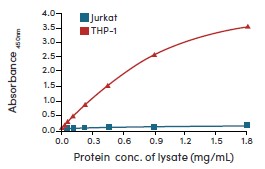
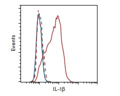
Cellular Readouts for Neuroinflammation• IL-1β/6/10
IL-1β (D3U3E) Rabbit mAb #12703: Flow |
Cell Death
Cell death serves as a readout for neurodegenerative disease progression. Neurons and glia can die and become diseased due to the aberrant regulation of cell death and survival pathways, which may include mitochondrial dysfunction, autophagy, and the activation of both apoptotic and non-apopototic pathways. With validated antibodies, protocols, and sampler kits from Cell Signaling Technology (CST), neurodegenerative disease progression can be reliably assessed by analyzing the different types and activation states of cell death in human and rodent samples.
Necroptosis• RIP, Phospho-RIP (Ser166)/(Ser14) |
Apoptosis• Caspase-3, Cleaved Caspase-3 (Asp175) |
Pyroptosis• Gasdermin D, Cleaved Gasdermin D (Asp275)/(Asp276) |
Proteinopathies
The production and processing of proteins that form protein aggregates are associated with frontotemporal lobar degeneration and taupathies. Therapies targeting protein aggregate formation may help slow proteinopathy progression. Protein aggregates found in blood or cerebrospinal fluid may also serve as biomarkers for diagnosing conditions earlier, and for monitoring disease progression and therapeutic response. Cell Signaling Technology® (CST®) antibodies are an ideal foundation for biomarker-based assays because they are thoroughly validated for specificity and sensitivity on biologically relevant binary model systems.
Amyloid• APP
β-Amyloid (D54D2) XP® Rabbit mAb #8243: |
Tau/Phospho-Tau• ELISA Kits
PathScan® Phospho-Tau (Thr217) Sandwich |
Additional Neurodegenerative Aggregate Markers• α-synuclein, Phospho-α-synuclein (Ser129)
Huntingtin (D7F7) XP® Rabbit mAb #5656: |
Cell Viability
Measuring cell viability can serve as a readout for determining neuronal disease progression. Aberrant or reduced proliferation, increases in neural damage, and the accumulation of senescent cells are all indicators of cell health and neurodegenerative disease status. CST offers antibodies and assay kits to determine cell viability efficiently and economically.
Cell Proliferation Markers• BrdU ELISA & Flow Kits
Ki-67 (D3B5) Rabbit mAb #9129: Confocal IF analysis of the ventricular zone in P21 mouse brain using #9129 (green). Actin filaments were labeled with DyLight™ 554 phalloidin #13054 (red) and DNA labeled with DRAQ5® #4084 (blue). |
Neural Damage Markers• Neurogranin
PathScan® Total Neurofilament-L Sandwich ELISA Kit #99175: Neurofilament-L protein is detectable in mouse and rat brain, but not HeLa cells (negative control) using #99175, as expected. |
Senescence• p16
Senescence β-Galactosidase Activity Assay Kit (Fluorescence, Plate-Based) #23833: |

Metabolism
The human brain is responsible for about 20% of basal energy expenditure, despite accounting for only 2% of body weight. Metabolic dysregulation and neurodegeneration are strongly correlated. Metabolic proteins may act as therapeutic targets since abnormal glucose tolerance or insulin resistance is observed in many neurodegenerative conditions. Cell Signaling Technology (CST) provides high-quality antibodies and assays to support the interrogation of metabolic pathways and cellular energy homeostasis regulation.
Metabolite Transporters• ABCA1• ABCA7 • ApoE4 • LRP1 |
|
|
Insulin Receptor Signaling• Insulin receptor |
Autophagy• mTOR |
Mitophagy• Pink1 |
Synaptic Plasticity
Synaptic plasticity is not only a part of development, learning, and memory, but has also been utilized as a readout for the clinical diagnosis of neurodegenerative diseases, including synaptopathies like Alzheimer’s disease. CST provides high-quality antibodies and assays to support the interrogation of synaptic activity and function.
Presynaptic Proteins• Synaptophysin
Synaptophysin (D8F6H) XP® Rabbit mAb #36406: Confocal IF analysis of mouse brain using #36406 (green), Neurofilament-L #2835 (yellow), Lamin A/C (4C11) Mouse #4777 (red), and DAPI #8961 (blue). |
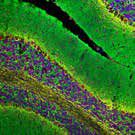
Postsynaptic Proteins• PSD95/Phospho-PSD95
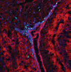
PSD95 (D27E11) XP® Rabbit mAb #3450: Confocal IF analysis of rat cerebellum using #3450 (red), Neurofilament-L #2835 (green), DRAQ5® #4084 (pseudocolor blue). |
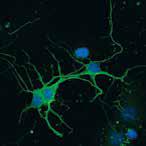
Modulators of Synaptic Plasticity• CaMKII
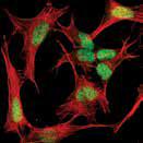
CaMKII-α (6G9) Mouse mAb #50049: Confocal IF analysis of primary neurons using CaMKII-α #50049 (green) and DRAQ5® #4084 (pseudocolor blue). |
Epigenetics
Brain health is heavily reliant on epigenetic mechanisms, and a loss of chromatin dynamics is observed in neurodegenerative diseases. Profiling epigenetic patterns and identifying the chromatin marks associated with disease progression have become increasingly important for the development of therapies against neurodegeneration. Cell Signaling Technology (CST) provides a comprehensive and diverse catalog of epigenetics products and an extensive assay portfolio used to measure protein-DNA interactions and histone modifications.
Histone Modifications• Acetyl-Histone H4 (Lys16)
|
DNA Methylation• 5-mC, 5-hmC
MeCP2 (D4F3) XP® Rabbit mAb #3456: Confocal IF analysis of SH-SY5Y cells using #3456 (green) and actin (red). |
Immediate Early Genes and Associated Proteins• CREB/Phospho-CREB
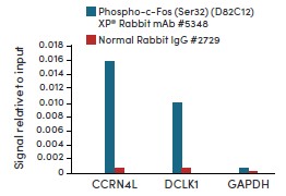
Phospho-c-Fos (Ser32) (D82C12) XP® Rabbit mAb #5348: qRT-PCR results for ChIP-enriched DNA from PC-12 cells starved overnight and treated with hβ-NGF #5221 and either #5348 or Normal Rabbit IgG #2729 using SimpleChIP® Enzymatic Chromatin IP Kit (Magnetic Beads) #9003. |
RNA-Binding Proteins (RBPs)
RBPs are increasingly becoming therapeutic targets for the treatment of neurodegeneration. Dysregulation of RNA-binding proteins (RBPs) has been linked to amyotrophic lateral sclerosis (ALS), Alzheimer’s disease, and frontotemporal dementia. Mutations in stress granule-associated RBPs lead to the pathological accumulation of protein aggregates that are readily apparent in neurodegenerative disorders. Age-related changes in mRNA methylation and methylation-dependent RBPs have also been associated with neurodegeneration. CST offers antibodies against key RBPs, stress granules, and m6A methylators, validated in relevant neuronal systems.
RBPs and Stress Granules• TDP43, FUS
TDP43 (D9R3L) Rabbit mAb (Alexa Fluor® 555 Conjugate) #68779: Confocal IF analysis of mouse cerebellum using #68779 (red), Ras (Alexa Fluor® 647 Conjugate) #37182 (cyan pseudocolor), and DAPI #8961 (blue). |
RNA m6A Methylation• N6-Methyladenosine
YTHDF2 (E2I2H) Rabbit mAb #71283: Confocal IF analysis of mouse medulla oblongata using #71283 (red), GFAP (Alexa Fluor® 488 Conjugate) #3655 (green), and DAPI #8961 (blue). |
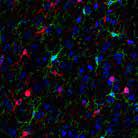
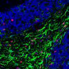
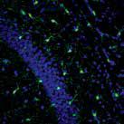
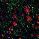
Multiplex Immunofluorescence (IF) Enablement
Cell Signaling Technology (CST) has the breadth of products available to enable several methods of multiplex staining, highlighted below.
|
|
|
|
|
|
Antibody Pairs for Platforms and ELISA
Rigorously validated antibody pairs are at the heart of every ELISA experiment. Cell Signaling Technology (CST) offers matched antibody pairs, both as a complete solution in sandwich ELISA kits or as custom pairs, ideal for incorporating into standard high-throughput ELISA-like assay platforms. These validated antibody pairs offer unrivaled specificity to deliver accurate readouts when monitoring therapeutic modulation of key neurodegenerative disease biomarkers. Some CST kits are sensitive to plasma proteins, enabling monitoring of disease status with live models.
Measure Key Biomarkers with Highly Specific and Sensitive ELISA Kits
|
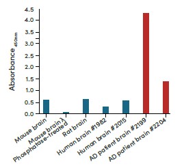
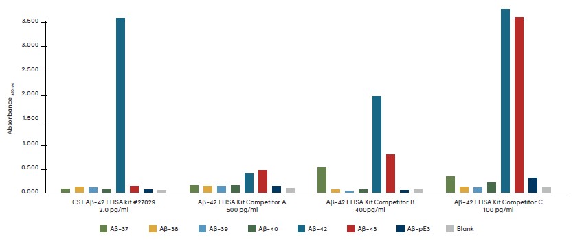
The PathScan® β-Amyloid (1-42) Sandwich ELISA Kit #27029 is highly specific to only human Aβ-42, showing no significant signal with Aβ-37, Aβ-38, Aβ-39, Aβ-40, Aβ-43, or pE3 peptides, as expected. By contrast, other leading commercially available β-amyloid (1-42) sandwich ELISA kits show little-to-no specificity towards the advertised Aβ-42 target. |
|
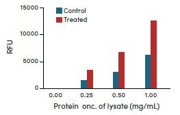
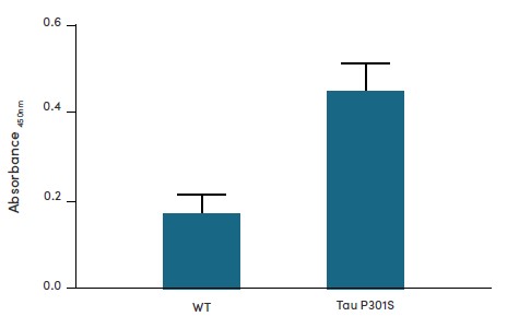
The FastScan™ Phospho-Tau (Thr181) ELISA Kit #58537 detects human tau in plasma from the hTau P301S mouse model in as little as 50 μL of plasma.
Data kindly provided by Dr. Li-Huei Tsai (Massachusetts Institute of Technology) and used with permission. |
Neuronal Markers
Studying the brain and nervous system requires examination not only of neurons, but also of microglia, oligodendrocytes, and astrocytes. The key to visualizing and identifying each of these cell types lies in using antibodies that target protein biomarkers specifically expressed and localized within these cells. Cell Signaling Technology (CST) provides highly specific, validated antibodies against accepted neuronal markers.
Immature Neuronal Markers• Doublecortin
Doublecortin (A8L1U) Rabbit mAb #14802: Confocal IF analysis of P5 mouse cerebellum using #14802 (green), GFAP #3670 (red), and DRAQ5® #4084 (psuedocolor blue). |
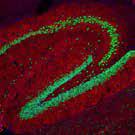
Mature Neuronal Markers• MAP2
NeuN (D4G4O) XP® Rabbit mAb #24307: Confocal IF analysis of mouse hippocampus using #24307 (green) and actin labeled with DyLight™ 554 Phalloidin #13054 (red). DRAQ5® #4084 (pseudocolor). |
Glial Markers
Glial cells consist of the non-neuronal supporting cells, including astrocytes, oligodendrocytes, and microglia. Distinct changes in glial cells and their markers are known to be associated with neurodegenerative disease progression. Glial cells also play an important role in overcoming challenges with the blood-brain barrier. CST has a wide variety of validated glial marker antibodies to identify and study the different glial cell types.
Astrocytes• GFAP
S100B (E7C3A) Rabbit mAb #90393: Confocal IF analysis of fixed frozen mouse cerebral cortex. #90393 (red), HS1 (Alexa Fluor® 488 Conjugate) #68206 (green), and DAPI #8961 (blue). |
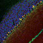
Oligodendrocytes• MBP
Olig2 (E6G6Q) XP® Rabbit mAb #65915: Confocal IF analysis of fixed frozen mouse cerebellum. #65915 (red), GFAP (Alexa Fluor® 488 Conjugate) #3655 (green), |
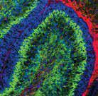
Microglia• Iba1
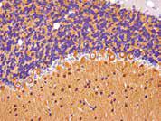
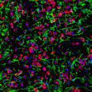
Iba1/AIF-1 (E4O4W) XP® Rabbit mAb #17198: Confocal IF analysis of mouse CA1 hippocampus using #17198 (green) and DAPI (blue). Images kindly provided by Dr. Marco Colonna (Washington University) and used with permission. |
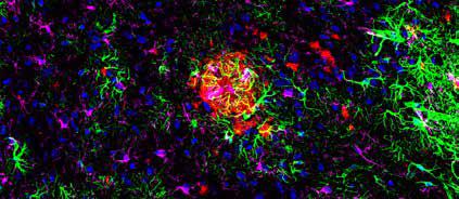
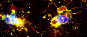
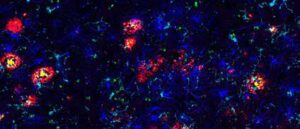
DAM Markers• ApoE
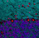
TMEM119 (E3E1O) Rabbit mAb #90840: Confocal IF analysis of an amyloid mouse model of Alzheimer’s disease brain using #90840 (green), GFAP #3670 (yellow), β-Amyloid (Alexa Fluor® 647 Conjugate) #42284 (red), and DAPI #8961 (blue). |